19 rue de Téhéran 75008 Paris  FRANCE

Dr Benoît PHILIPPE

Maxillofacial and Oral Surgeon

19 rue de Téhéran 75008 Paris  FRANCE
style4_homepage

Dental implants

Dental implants are artificial roots made out of titanium and made to support dental prostheses (crown, bridge, etc.). In the absence of contraindication, dental implants can replace the missing tooth in case of edentulism.

The dental implant procedure is a surgical act. Therefore, it requires a pre-surgical assessment and a preliminary study of the surgery feasability according to data acquired through science, mandatory human and technical means (surgical equipment, operation room, sterilization of the equipment and traceability meeting the standards in force).

The main requirements to a successful and long-term dental implant stability are to stop smoking, to have good dental care before and after the implant placement (maintenance), to abide by contraindications (especially biphosphonates, radiotherapy and alcohol), and have a thick attached gum around the implants.


  1.     Pre-surgery assessment and feasability study


The pre-surgery assessment defines the patient’s needs, seeks possible contraindication and presents the whole information necessary to an informed consent (surgical procedure, type of anaesthesia required, surgery follow-up and possible complications, estimate).

The pre-surgical assessment is run by the practitioner in charge of the dental prosthesis. During the pre-surgical assessment, the number and precise location of the implants, their shape, their length, their respective diameter etc. will be determined.

The assessment steps are the following :

  1.    Set up the radiograph guide. It is elaborated by the technician of the practitioner in charge of the prosthesis (crown, bridge, etc.) or by specialized softwares. The radiograph guide accurately reproduces the future dental prostheses. As it is radio opaque (i.e visible during the radiograph assessment) the surgeon can assess the quality and the volume of the bone available to the implementation of the dental prosthesis (i.e the location where the dental implants will be placed).
8
8
6
6

2.    Perform a radiograph assessment (scan, Cone Beam). It confirms the bone volume available (quantity and quality study) and tests the placement of implants thanks to specialized softwares.

9
9
10
10

3.  Set up the surgical guide. It is made by the technician of the prosthodontist thanks to the radiograph guide or on a 3D printer thanks to computer-aided simulation. It controls the surgical procedure thus ensuring maximum accuracy and safety.

11
11
12
12

Surgical Procedure  


Most of the time dental implants are placed between 2 to 12 months after tooth extractions on a healed bone. However, implants can sometimes be placed on the same day as the tooth extractions. The benefit of this technique is to shorten the whole treatment duration but it requires for the surgeon to have access to a surgical extraction site free from any infection as well as an adequate anchoring for the implants in the extraction site.

The implant procedure is conventionally divided into two stages.

Stage I : It consists in placing the implants in the bone. The implants are embedded in the bone for two to four months.

Stage II : It consists in setting healing screws through the gum. Healing screws maintain the gum open during the construction phase of the prosthesis on the implant. After the gingival healing period lasting around two weeks, the dental prosthesis practitioner places the crowns or bridges on the implants.

13
13
14
14
15
15
37
37
16
16
18
18
17
17
19
19
20
20
21
21
final
final

Other possible procedures :    

  • Immediate loading procedure. The dental prosthesis is placed on the same day as the implant placement. This procedure is particularly attractive as it shortens the treatment duration and suppresses the discomfort of edentulism. However, a good quality bone is paramount and a precise control of chewing forces. Temporary prosthesis are necessary in immediate loading procedure.


  • Placing implants with an abutment through the gum on which the dental prosthesis is fastened (Tissue Level Technique). The benefits of this procedure are to shorten the rehabilitation duration such as reducing the number of surgical acts and gingival trauma but it requires to anticipate the length of gingival healing.
24
24
25
25

Pre-implant Surgery


Pre-implant surgery refers to all surgical acts enabling to deal with implant contraindications.

The most frequent contraindications against dental implant placement are the lack of bone volume ( skeletal atrophy) and the incorrect relation between the two toothless jaws (upper maxilla and lower mandible).

Secondary bone loss (or acquired bone loss) can be due to :

  • Physiological ageing of the jaw with edentulism (normal ageing linked to the age of the patient)
  • « Pathological ageing » of the jaw (i.e. premature ageing) notably for patients suffering from chronic periodontitis. Smoking and poor dental hygiene are the most frequent triggering factors.
26
26
27
27
28
28
  • Sequela after a trauma (jaw fracture with bone fragment loss)
  • Following a tumor surgery (resection of a more or less extensive fragment of the jaw bone whether it be a benign tumor or a malignant tumor of the maxilla
  • Failure of previous implant surgical acts (graft failure or implant failure)
29
29
30
30
31
31

Primitive bone loss. Primitive bone loss is scarcer. Several types of primitive bone loss are described :

  • Simple primitive bone loss : the patient has a slender morphotype. The jaw bones are too thin and dental implants can’t be placed.
  • Primitive bone loss due to an absence of teeth (single or multiple dental agenesis). It is a congenital condition (birth defect).
  • Primitive bone loss due to congenital bone deformities : facial clefts and other syndroms.
32
32
33
33
34
34
  1. Reconstruction Techniques


The following aspects are taken into account

  • Height increase of the bone volume
  • Thickness increase
  • Combined height and thickness increase


These increases can affect either the maxilla (upper jaw) or the mandible (lower jaw) or both. Similarly, these increases can affect the dental arch in full or partly.

Bone loss is often combined with gum atrophy ( loss of attached gingiva). A reconstruction of the attached gingiva is mandatory to ensure a long lasting result to the implant rehabilitation.

atrophie-osseuse-manque-de-hauteur
augmentation-de-hauteur
7-ans-apres-implantation
  1. Choice of the reconstruction material : Autogenous bone, Bone substitute, or Biomaterials?


Various materials can be used to rebuild the atrophied jaw :

  • Autogenous bone : it is harvested from the patient’s own body on the day of jaw bone reconstruction.


  • Allogenous bone : it comes from another human being’s body (Bank-bone)


  • Xenogenous bone : it comes from other species (i.e animals, particularly bovine species)


  • Bone substitute biomaterial of natural or synthetic origin.


The Choice of the grafting material depends on numerous factors :

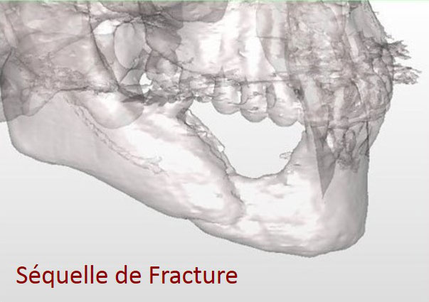
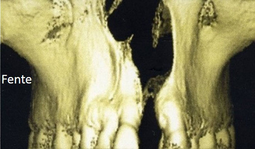
  • The location, size and shape of the bone atrophy
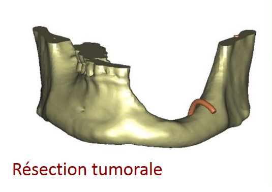
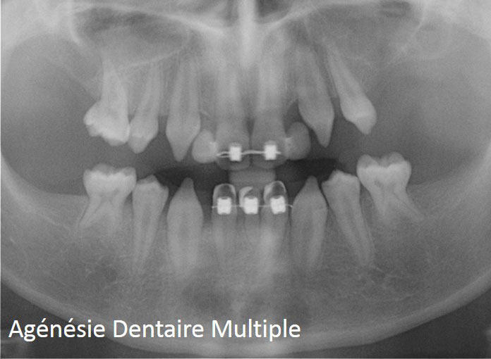
  • The cause of the atrophy : ageing, periodontal disease, trauma, birth defect, tumor sequela….
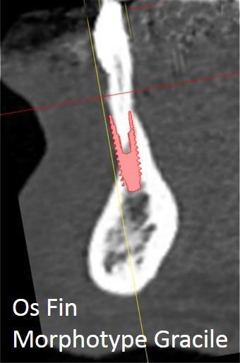
  • The quality of the gum tissue
  • The patient’s medical profile (age, medical and surgical history…)
  • The number and position planned for the implants.


Even though bone harvesting is required when performing autogenous bone grafting thus involving an extra surgical room, this procedure is still highly beneficial :

  • The autogenous bone is harvested on the patient’s own body, therefore avoiding any infectious disease transmission (bacterial, virus, prion) and any immune mediated rejection.
  • It is alive and already calcified during harvesting.


The autogenous bone is mostly used particularly in case of widespread atrophy.

There are numerous autogenous donor sites available. The reasons leading to the choice of a particular donor site or sites depend on the needs required for the reconstruction (quantity required, density, shape, etc.) and are explained thoroughly to the patient during the pre-operative consultations (informed consent).

Bone substitutes and biomaterials are beneficial as they avoid bone harvesting. They are most of the time used for small volume reconstructions or in addition to the autogenous bone in case of a large reconstruction. Bone substitutes authorized by the current French and European legislation show no risk of transmission of infectious disease (bacterial, virus, prion) or immune mediated rejection.

Whatever the reconstruction or material used, the patient will be explained thoroughly the criteria leading to the choice of procedure during the pre-operative consultation (informed consent)

  1. Schedule. General treatment scheduling


Whatever the reconstruction procedure chosen, the steps in case of reconstruction are the following :

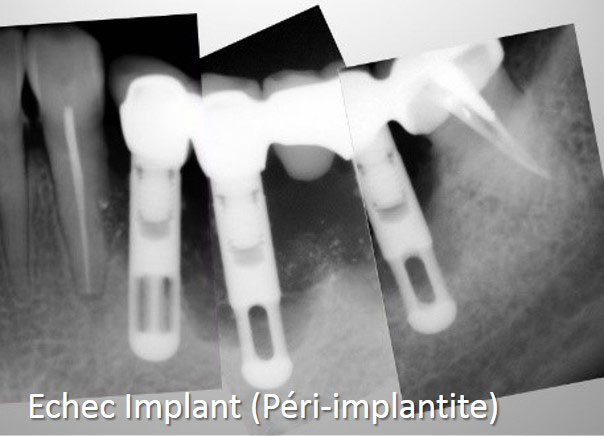
  1. Suppress any source of infection which may lead to reconstruction failure (removal of any remaining broken tooth or infected tooth, thorough dental scaling)
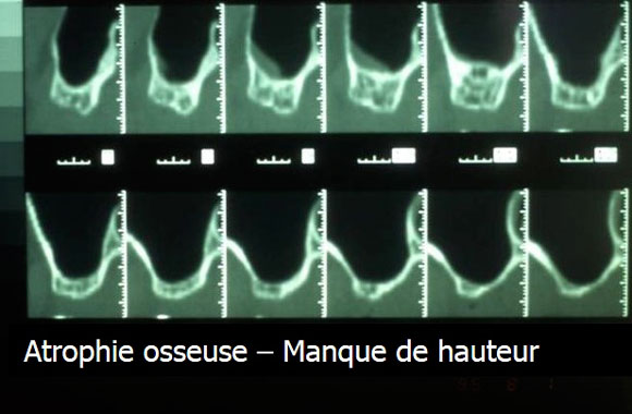
  2. Perform an X-ray in order to assess the bone loss and choose the most suitable type of reconstruction (height increase, thickness increase, mixed increase).
  3. Rebuild the bone volume
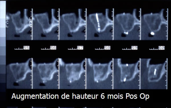
  4. Comply with a bone healing time of 5 to 6 months. During this step the patient may wear a removable denture provided it doesn’t compress the reconstructed area.
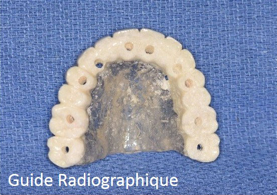
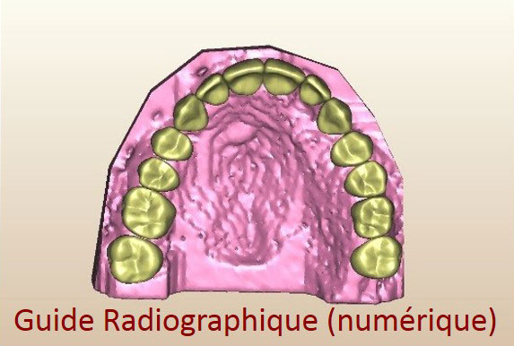
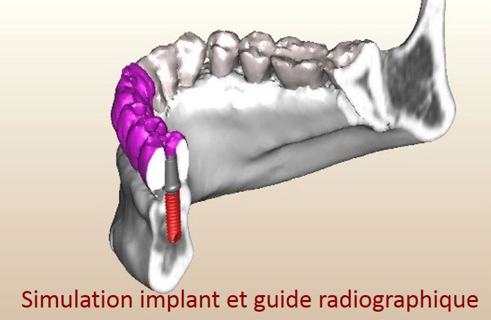
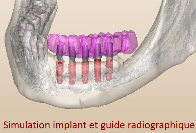
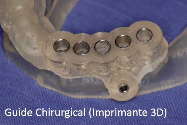
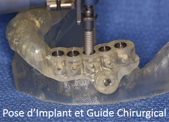
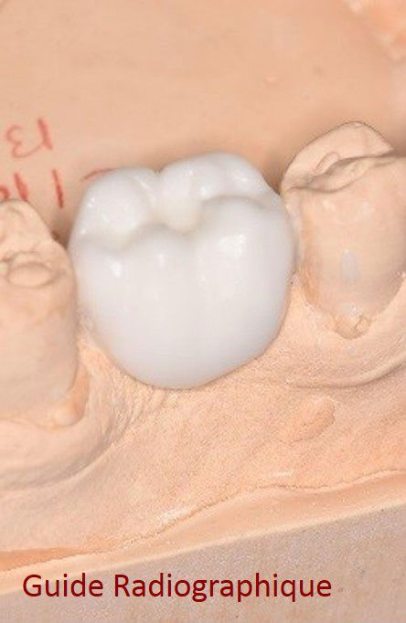
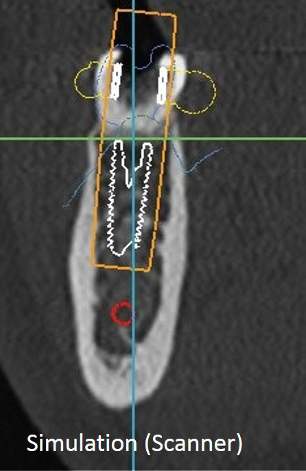
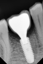
  5. Perform an Cone-Bean CT or CT Scan after the healing period to confirm the reconstruction in order to check the quality of the reconstruction and simulate the placement of the implants. The patient has a radiograph guide during the exam.
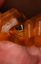
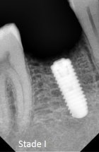
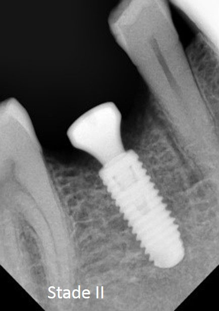
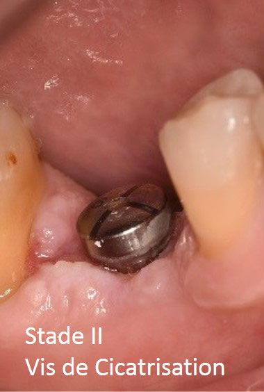
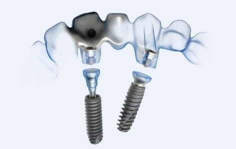
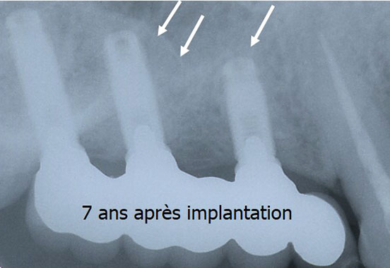
  6. Dental implant surgery
  7. Comply with an implant healing time of 2 to 6 months. During this step the patient may wear a removable denture provided it doesn’t compress the gengiva regarding the implants.
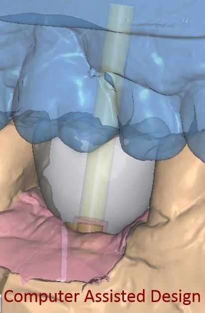
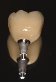
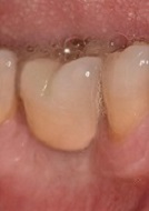
  8. Manufacture the dental prosthesis. Nowadays, to benefit from a greater accuracy to adapt dental prostheses to the underlying implants, the infrastructure of dental prostheses (crowns, bridges, etc.) is realized more and more thanks to computer softwares (CAD-CAM) (see videos).


  • Other Procedures and Associated Procedures


Osteotomies


Osteotomies are designed to correct the abnormal distance between the two jaws. It is especially the case when edentulism is combined with alveolar bone loss. The space between the two jaws is too wide and prevents the production of a satisfactory implant prosthesis on a bio-mechanical level (the implants are too tilted or too short) or on a aesthetic level (a simulated gum tissue looks unsightly)

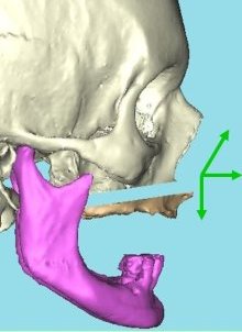
As in the jaw malformation surgery (see maxilla surgery or orthognathic surgery) osteotomies can deal with the whole skeleton of the jaw (complete osteotomies) or only one segment (segmental osteotomies).

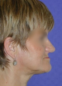
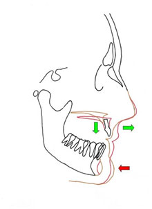
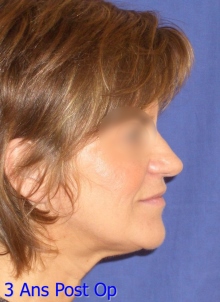
The most frequent case is bone loss which is combined with complete edentulism of the upper maxilla. The resorbed toothless maxilla is located too far back compared to the mandible while the upper lip is set back as it is not supported. The distance between the upper maxilla and the lower maxilla is too wide. The surgical act consists of performing a Lefort 1 complete osteotomy moving the atrophied maxilla forward and downward while performing simultaneously a reconstruction of the bone structure using autogenous bones of iliac origin. This bone graft is placed in the intermediate space newly created by the forward and downward movement of the maxilla. Most of the time, onlay bone graft using autogenous bone of calvaria origin is performed jointly in order to thicken the maxillary crest which was too thin due to bone resorption phenomena.

Cube1a
Cube1a
Cube1b
Cube1b
39
39
Cube1c
Cube1c

More scarcely, when facing a local bone atrophy, only one segment of the crest may be moved (segmental osteotomy) in order to recreate a satisfactory prosthetic space both on an aesthetic level and mechanical one.

Gingival grafts


In addition to a proper dental hygiene (brushing one’s teeth after eating, periodic and consistent scaling, stopping smoking), having a thick gingiva attached to the alveolar bone around the implants is a prerequisite for a long lasting result.

Therefore, in case of gingival atrophy, the gum tissue must be rebuilt. Numerous techniques exist to solve this problem ( gingival tissue graft harvested on the palate, graft of connective tissue, etc.) Gingival grafts can be performed before or after the placement of implants. In any case, it is better to have them performed by a specialist (Periodontologist).