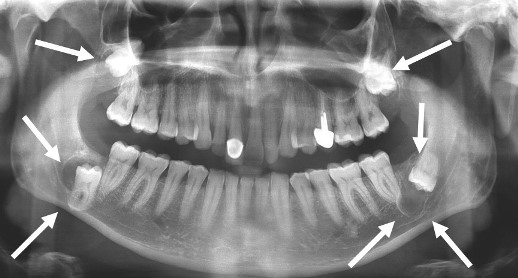
The third molars (wisdom teeth) are the most common instances of retained or impacted teeth found in the maxilla and / or the mandible.
However, all other teeth especially canines may be retained or impacted.
The third molars (wisdom teeth) are the most common instances of retained or impacted teeth found in the maxilla and / or the mandible.
However, all other teeth especially canines may be retained or impacted.
The surgical indications of retained or impacted teeth are based on objective signs. They can be :
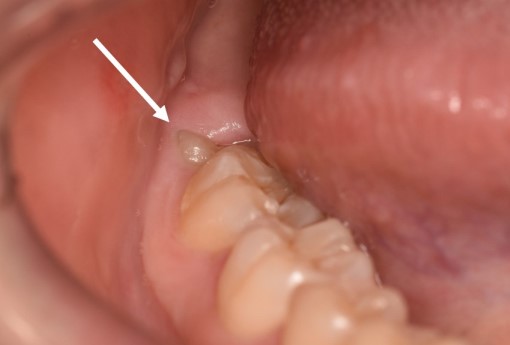
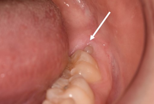
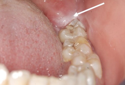
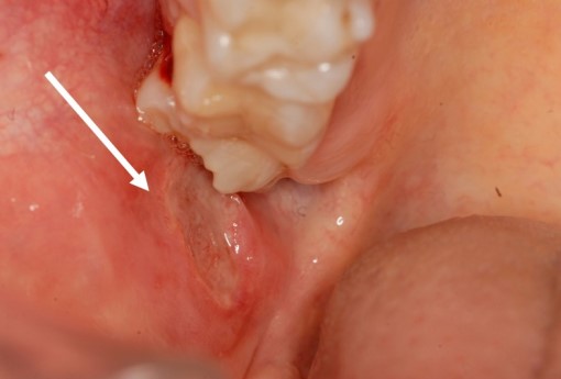
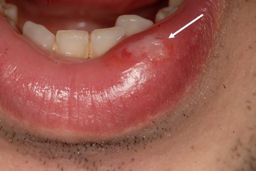
The medical exam shows a tooth fragment which seems to be erupting from the gum in the back of the mouth. The gum is swollen, painful, inflamed and even purulent, more or less ulcerated. It is therefore referred to « disimpaction signs».
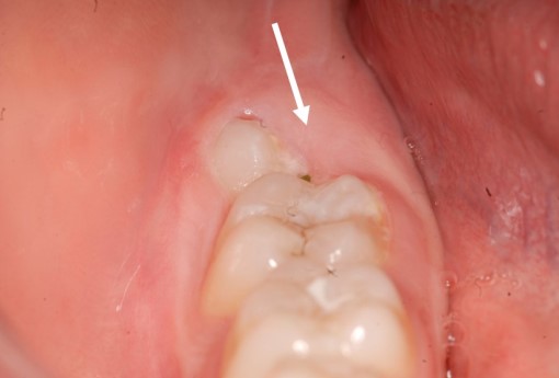
Disimpaction signs are more often located in the lower jaw (mandible) than in the upper jaw (maxilla). When an upper wisdom tooth has a tilted eruption towards the cheek due to a lack of space on the dental arch, a painful ulceration of the cheek is often observed. Difficulty to open the mouth often happens alongside this type of ulceration.
Wisdom teeth infections may be limited locally but they can also spread quickly to the whole face and neck thus leading to an emergency (neck cellulitis) requiring an emergency admittance (high dose of antibiotic therapy, medical supervision followed by tooth extraction as soon as opening the mouth is possible). Non-steroidal anti-inflammatory drug (NSAID) are absolutely contraindicated in case of proven infection. Patients must contact their physician as soon as possible and must NOT self-medicate with such NSAID
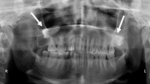
Au maxillaire supérieur, une radio panoramique dentaire systématique peut révéler l’existence de dents de sagesse incluses totalement silencieuses et pour lesquelles l’extraction s’avère nécessaire avant l’apparition de complications (sinusite, troubles ophtalmologiques, douleurs invalidantes de la région de l’oreille, névralgies faciales, etc.)
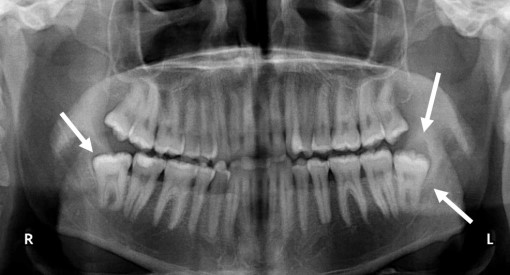
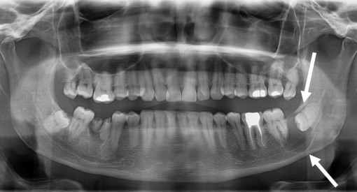
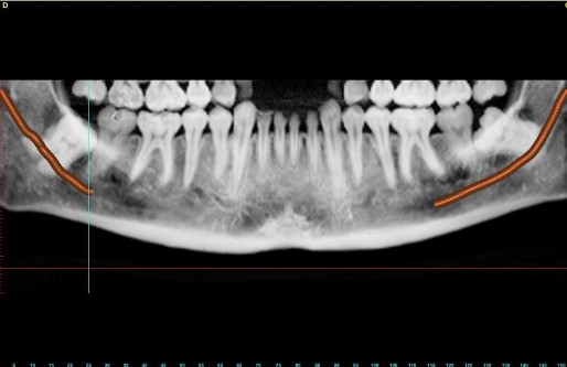
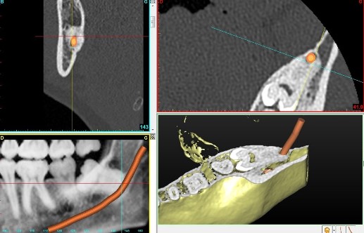
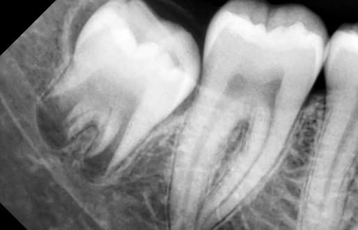
In adults, wisdom teeth can be close to the inferior dental nerve because their roots have grown. The dental nerve is responsible for the sensitivity of the lower teeth as well as the sensitivity of the lower lip and chin. If the panoramic dental X-ray shows that the tips of the roots are too close to the dental nerve then a Cone Beam CT is always prescribed so as to assess the risk of damaging the nerve during extraction. Therefore, the surgeon can know before surgery the exact position of the nerve compared to the roots (forward, backward, in between). In any case and despite all the measures taken, the patient is warned about a possible nerve damage during the preoperative appointment (informed consent).
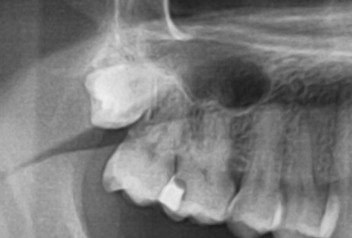
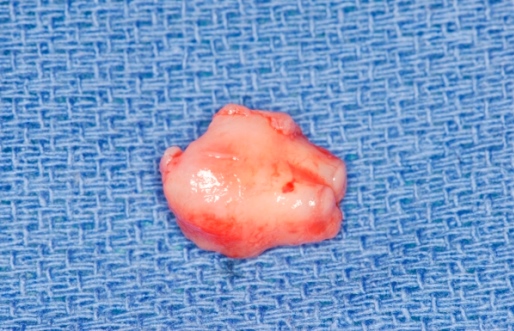
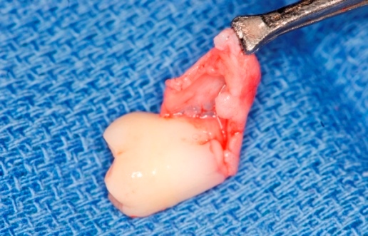
The roots of the wisdom teeth in their germ state have not developed yet ; they are referred to « wisdom teeth germs ». Only the developed and mineralized crown surrounded by a the pericoronal bag tissue is visible during X-ray.
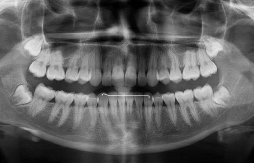
In case of an orthodontic treatment and when there is an obvious lack of space on the dental arch, it is better not to wait for the end of the growth to have the wisdom teeth extracted. It is indeed easier to remove the germs without any roots (germectomy) than to remove grown teeth with long roots retentive to the bone and in close contact with the dental nerve. The surgical follow-up is also more simple as germs without roots are easier to extract and surgery scarcely causes any nerve damage.
Even though the comfort and safety of the patient is a priority, in case of hospitalization, surgery of impacted or retained wisdom teeth requires outpatient surgery a.k.aambulatory surgery or same-day surgery (hospitalization is only a few hours). Hospital discharge is under the authority of the surgeon and anesthesiologist.
Several types of exams are available. They can be :
1. Local care. Applying natural or synthetic ice to reduce swelling, pain and inflammation
2. Prescription drug.
3. Diet
Several post-surgical medical considerations are taken into account concerning the patient’s diet and modify it (edema of the cheek and lips, numbness of the lips, the patient’s fear to chew after a recent jaw surgery, etc.)
For further information, please contact us. Dr Benoît Philippe’s secretariat is available to assist patients and answer any additional query by phone 0147429070 or by email